Natural Farming

Natural Farming

Soil - our connector!

Soil - our connector!
Showing posts with label Diseases. Show all posts

Saturday, January 25, 2020

Rift Valley Fever (RVF)


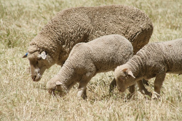
RVF is a mosquito-borne zoonotic viral disease affecting many animals, especially domestic livestock, and humans. It is most common in eastern and southern Africa. Sheep, in particular exotic, introduced breeds, are most susceptible.

It can cause fever, haemorrhage and abortion in pregnant animals. More than 90% of infected lambs die; mortality of adult sheep is as low as 10%. Humans can contract RVF from mosquitoes and possibly other bloodsucking insects.
RVF epidemics have a serious impact on the economy due to death of livestock, infection of humans and bans on livestock trade from RVF-infected areas.

Rift Valley Fever  - symptoms and prevention
Is an infectious zoonotic disease affecting sheep, goats, and cattle.
Rift Valley Fever (infectious enzootic hepatitis) and humans are susceptible to the  disease.
Rift valley disease is a viral disease of sub-Saharan Africa. The virus attacks the liver and causes symptoms ranging from fevers and listlessness to hemorrhage and abortion rates approaching 100% in pregnant sheep. It is transmitted by mosquitos. RVF is a notifiable disease and it is thus important for farmers to inform the State Vet and Animal Health Technician when they suspect the disease.
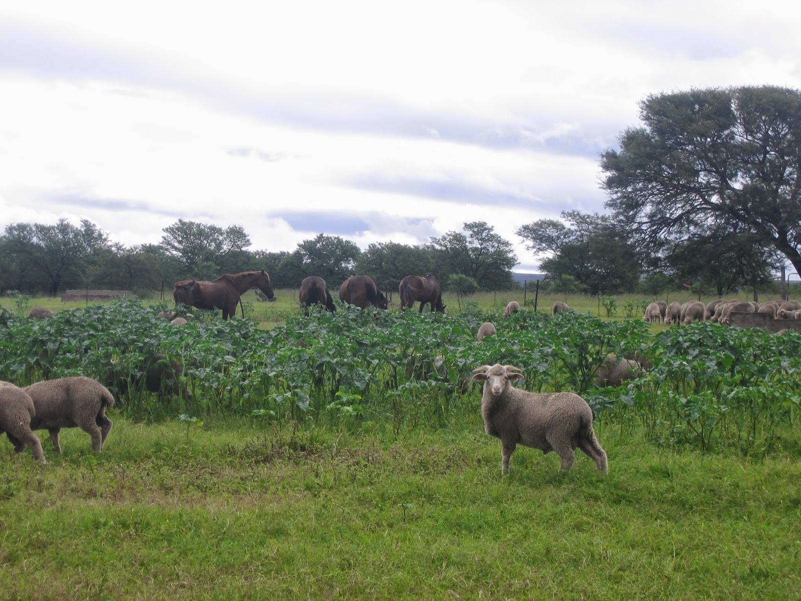
Vaccination of animals against RVF has been used to prevent disease in endemic areas and to control epizootics. Rift Valley fever is more deadly than West Nile virus. Animals should be moving away from standing water and moved to higher altitude areas. Caution must be exercised when handling infected tissues by wearing gloves, masks, goggles and by using viricidal disinfectant. The incubation period is 1 – 3 days.
Symptoms in animals:
Abortions, mortalities in young animals, jaundice, lagging behind, weakness and exhaustion, bloody diarrhea, bleeding from the nose and fever.
Symptoms in humans:
Influenza-like symptoms – headaches, muscle pain, joint pain, abdominal pain and nausea.
Prevention
Vaccinate sheep older than 6 months. Inactivated vaccine can be used in pregnant ewes. Animals must be given a booster within 3-4 weeks after initial vaccinations and then must be done annually.
Movement restrictions are recommended. No movement without notifying the Provincial State Vet.The disease is caused by the Rift Valley Fever (RVF) virus, a member of the genus Phlebovirus in the family Bunyaviridae and the disease is transmitted by mosquitoes.Limited to Africa in earlier years, it causes the enormous waste of livestock, especially in wet conditions
For decades Rift Valley fever has caused the illness and death of large numbers of livestock in Kenya and in much of sub-Saharan Africa. Increasing evidence linking the disease to human deaths as well has led epidemiologists to include Rift Valley fever on the list of emerging viruses (including HIV and Ebola) that infect thousands of people each year.
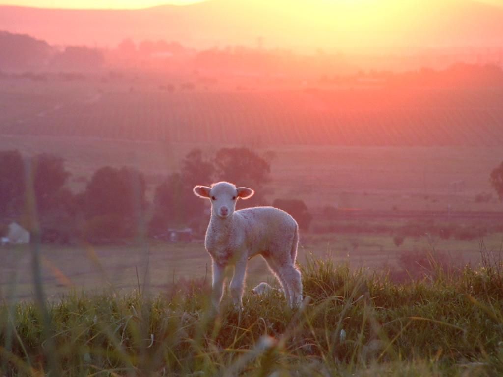
Clinical Signs
In young lambs the incubation period varies from 20 to 72 hours. Some lambs die suddenly without showing signs of this disease. Usually, however, affected lambs develop fever, refuse food, physically weaken, recline and die after a course of 24 hours. Mortality often reaches 95%.
In adult sheep the most common clinical finding is abortion. Most affected sheep show fever of 41 to 42 °C, abortion and vomiting. During fever, severe leukopenia, especially of neutrophils, forms.                

Controlling RVF in animals
•           Outbreaks of RVF in animals can be prevented by a sustained programme of animal vaccination. Both modified live attenuated virus and inactivated virus vaccines have been developed for veterinary use. Only one dose of the live vaccine is required to provide long-term immunity but the vaccine that is currently in use may result in spontaneous abortion if given to pregnant animals. The inactivated virus vaccine does not have this side effect, but multiple doses are required in order to provide protection which may prove problematic in endemic areas.
•           Animal immunization must be implemented prior to an outbreak if an epizootic is to be prevented. Once an outbreak has occurred animal vaccination should NOT be implemented because there is a high risk of intensifying the outbreak. During mass animal vaccination campaigns, animal health workers may, inadvertently, transmit the virus through the use of multi-dose vials and the re-use of needles and syringes. If some of the animals in the herd are already infected and viraemic (although not yet displaying obvious signs of illness), the virus will be transmitted among the herd, and the outbreak will be amplified.
•           Restricting or banning the movement of livestock may be effective in slowing the expansion of the virus from infected to uninfected areas.

•           As outbreaks of RVF in animals precede human cases, the establishment of an active animal health surveillance system to detect new cases is essential in providing early warning for veterinary and human public health authorities.

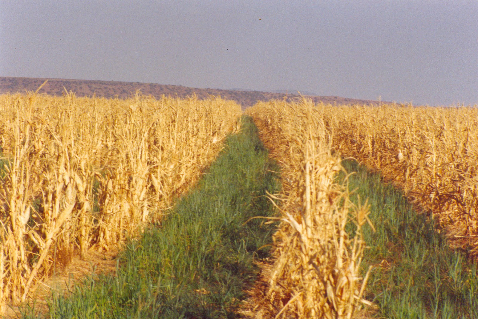
RVF FORESCASTING AND CLIMATIC MODELS
Forecasting can predict climatic conditions that are frequently associated with an increased risk of outbreaks, and may improve disease control. In Africa, Saudi Arabia and Yemen RVF outbreaks are closely associated with periods of above-average rainfall. The response of vegetation to increased levels of rainfall can be easily measured and monitored by Remote Sensing Satellite Imagery. In addition RVF outbreaks in East Africa are closely associated with the heavy rainfall that occurs during the warm phase of the El Niño/Southern Oscillation (ENSO) phenomenon.


Saturday, March 21, 2015

Heartwater infectious disease


General info with focus on South Africa. 


Heartwater is an acute infectious disease responsible for severe losses among susceptible cattle, sheep and goats. Its causal organism, Cowdria ruminantium, is transmitted by the bont tick, Amblyomma hebraeum. In nature the disease can be transmitted only by this tick in its nymphal and adult stages and occurs only where this tick is present.


The bont tick prefers warm and moist lowveld and bushveld areas and is found in Mpumalanga, Kwa-Zulu-Natal, the Northern and Northwest Provinces, Eastern and Western Cape Provinces, as far south as Mossel Bay. It also occurs in parts of Swaziland and Botswana.
Bont tick distribution in South Africa.


Affected animals have a fever of between 40 and 42 °C, are listless, loose their appetite and lag behind the rest of the herd. As the disease progresses, movement becomes more impeded. Animals often develop a high-stepping gait. Breathing becomes more labored and animals may push their heads up against firm objects. The affected animals eventually lie on their side with the head pulled backwards and the legs extended. They are sensitive to intensive light and twitch their eyelids rapidly when exposed to it. The eyes roll from side to side in the orbita. The body temperature rapidly drops to below normal prior to death.


A postmortem examination reveals varying quantities of straw-colored fluid, which may sometimes be blood-tinged, in the chest cavity and the heart sac (hence the name of the disease). The lungs are heavy and have a soaked appearance. White foam is often abundant in the air passages. Some or all of these changes may, however, also be observed in animals dying from other causes and only a veterinarian can make a final diagnosis by microscopical examination of a smear prepared from brain tissue.

Heartwater occurs throughout the year. The incidence varies according to the tick population and its activity and the disease is therefore less frequently encountered during winter. The disease occurs when an infected tick feeds on a susceptible animal. Bont ticks become infected with the heartwater organism when they feed on an infected animal in which the organism is circulating in the blood at that particular time. Engorged larval and nymphal ticks drop from the host, moult within 4 to 6 weeks and are ready to seek new hosts on which they can feed as nymphae and adults, respectively. If such a host is susceptible to heartwater they will transmit the infection to it. Infected adult female ticks play no further role after having engorged, dropped and laid their eggs, because they then die and the heartwater organisms are not transmitted through the eggs to the next generation of ticks. Only 2 to 5 % of bont ticks found in heartwater areas are infected.


The movement of animals carrying infected ticks into or through an area free from the disease, can result in losses among susceptible stock if they are infected by ticks which drop from the infested animals. The ticks, however, seldom survive longer than one season in an area which is climatically unsuitable for them.


Apart from cattle and small stock, large and small game animals, hares and some ground dwelling birds (eg guineafowl) can also become subclinically infected with heartwater, and although they may show no clinical signs of the disease, they may have the organism circulating in their blood and therefore be able to infect ticks that feed on them. As healthy carriers of the infection, these animals and birds play an important role in the maintenance of the infection in ticks and consequently also in the occurrence of the disease. Because they cannot be dipped like cattle, these animals and birds also regularly serve as hosts on which the ticks can feed. It is clear that the bont tick, and therefore heartwater, cannot readily be exterminated on a farm where large and small game are present.


Cattle, particularly indigenous breeds, older than 1 year and newborn calves up to an age of 4 to 6 weeks, have nonspecific resistance to the disease and when infected many of them show no clinical signs, or at most develop a mild fever reaction.

Heartwater organisms seen in a brain smear (arrow).
They are magnified approx. 1000 times!
Cattle and small stock may be resistant to infection by infected ticks for two reasons: animals may be immune (either as a result of an earlier infection or through vaccination) or they may have a varying degree of natural resistance against the disease. Animals born in a heartwater area are usually, but not necessarily always, exposed to infected ticks (and become infected) when they are still young and have an inborn natural resistance. In this way they acquire an early immunity, which is thereafter boosted by regular reinfection by ticks. 
Losses as a result of heartwater most often occur when animals born and reared in areas free from the tick and the disease are moved into areas where the disease occurs (endemic areas) or when animals in endemic areas loose their immunity, because they are not exposed to reinfection by ticks (eg as a result of a too intensive dipping program).

Prevention
Ruminants younger than three weeks of age have a natural resistance to heartwater infection. This resistance does not depend on colostrum intake. When a young animal is bitten by an infected tick, it will build up an immunity, but not become ill. If. however, it is bitten for the first time when it is older than three weeks, it will fall ill and may die if not treated appropriately. Animals that are immune have to be bitten at regular intervals since they may lose their immunity if not bitten within six to nine months. It is thus of the utmost importance that animals in heartwater endemic areas (areas where it occurs naturally) should not be kept free from ticks for extended periods.

Treatment
There are numerous drugs on the market containing Oxytetracycline as the active substance. All of these drugs will be effective against the heartwater organism provided it is used timeously and a high enough dose is given. The earlier treatment is begun the higher the chances of success.
In severe cases, cortisone may be given with the Oxytetracycline. These drugs, however, have serious possible side-effects and are only available on prescription from a vet. If given to pregnant animals, abortion may ensue. Cortisone helps to stabilize the damaged blood vessels to prevent further leakage of fluid and also aids in relieving excess pressure on the brain.

Monday, May 19, 2014

Pneumonia infections in sheep


Pneumonia infections in sheep may present in some cases as chronic or acute symptoms resulting in
death.
More often, young and unvaccinated sheep that have not been vaccinated for some period, may become infected. Poor hygiene conditions may often also have an additional impact in this condition. 

Certain conditions e.g. weaning, transportation, exposure to infected animals in feedlot conditions may cause stress and result in “losses” due to this disease complex.

Diagnosis:
Generally, diagnosis is conducted when deaths occur. A veterinary conducted post mortem may indicate a case of pneumonia. Ideally samples should also be sent to a veterinary pathology laboratory. Diagnosis will often be made with bacterial cultures, tissue sectioning for histopathology and circumstantial history.

Prevention:
The vaccination of all sheep of the correct “responsive” age, approximately 2-3 months should be
conducted.

Strategic Dosing - Sheep and Goats


As winter approaches in South Africa many farmers believe that the worm problem associated with the humid and often hot summer months will be drastically reduced with the decreasing temperatures. 
While this may be true, winter is not without its worm challenges. This said, flukes (conical and liver flukes), roundworms (Brown stomach worm, Bankrupt worm (Long necked), White bankrupt worm, over wintering roundworm and nasal bots larvae’s are some of the worm burdens your stock has to deal with during the winter period.

 There is increasing recognition that larvae arrest in development as a result of prior experience of certain climatic or seasonal influence, a phenomenon referred to as hypobiosis.  Low or zero egg counts in winter is therefore no assurance that there exists a low or no worm burden. These hypobiotic larvae will continue from their arrested development when favourable environmental conditions ensue:

     ·         During the next spring or,
    ·      When host (ewe) immunity around the next lambing time is reduced – a “normal” occurring phenomenon that we need to manage. 

Winter is also a critical time for ewes, most of whom are pregnant during a period often characterised by poor grazing quality and harsh weather conditions. Ewes are required to maintain and grow the foetus inside them – especially during the last third of pregnancy. Closer to term, there is an increased nutrient requirement towards formation of colostrum. An increased winter worm burden will, despite improved nutrition, erode on the ewe’s body condition (fat reserves), increase the risk of poor oöcyte (egg) quality and conception, decrease the quality of colostrum and put the newborn at risk. Additionally, anaemia, poor growth and diarrhoea are also seen in winter due to worm problems.

The fact pointed out above should make “pre-winter strategic dosing” an essential component of any farm’s integrated parasite management (IPM) system. Farms applying a “before winter strategic dosing” dose all their animals usually in May, after the frost, when there is little or no re-infection of the animals from the pasture. 

Using the correct remedy, pre-winter strategic dosing should achieve the following:


  • Eliminate nasal bot larvae in the sheep when there are no longer free living flies around that lead to re-infestation. Pre-lambing dosing against nasal bots is important as we need to ensure that the ewe can identify her lamb after birth by olfactory means. Similarly, rams are more efficient in detecting ewes in oestrus when nasal cavities are free of bots and the mucus secretion this infestation causes    
  • Remove hypobiotic roundworm larvae (positive egg count is therefore not a requirement for dosing).  

General Pathology:


Animal well-being is a priority for farmers and if one of the flock falls ill there is a best practice to follow. Here are some guidelines on what to do when things go wrong, to protect the rest of the flock. 

What you should keep in mind and do when you have a sick animal, before calling the vet for advice. 


General Pathology:

1.    In the living animal look for clinical signs. General examination – start at the head, remember feet and mouth (blue tongue); openings (discharges, colour, swellings, diarrhoea); mucous membranes, behaviour, temperature; general body condition;
2.    History!!!! – Clinical signs, vaccinations and previous treatments, diet, age group, veld type etc. are important.


Post Mortem:

  1. Animal must lie on it’s right hand side, lift forelimb and hind limbs and cut – look for subcutaneous lesions (bruising; anaemia).
  2. Cut abdominal cavity open gently along the midline – note if fluid comes out, and colour of fluid. Cut up to the end of the rib cage. Cut attachment of the diaphragm to the rib cage – listen for a popping sound – lugs collapsing due to loss of negative pressure. If the lungs do not collapse it could be a possible pneumonia/
  3. Cut open the thorax along the sternum so all the organs in the chest are exposed.
  4. Remove organs of the abdominal cavity: spleen (sample for bluetongue); gastrointestinal tract (parasites; bacterial infections; faecal collections); rumen contents (acidosis; plant remnants) mesenteric lymph nodes and ileocaecal lymph nodes (Johnes disease); liver (liver fluke; toxic plants; copper deficiency/toxicity; domsiekte); kidneys (pulpy kidney, copper toxicity). Look at the bladder and if full look at urine (possible copper toxicity if urine is red). Remove all organs carefully and only once samples have been taken cut into the organs.
  5. Do the same for the thorax. Cut open the trachea into the bronchi and into lung tissue. Cut open the heart (look at clot in ventricle).
  6. Head and Brain – brain can be sent in for histopathology if nervous symptoms have occurred e.g. thiamine deficiency.

Sample Collection – label them clearly!!!!


·         Bacteriology – as sterile as possible; all affected organs; tied off GIT loops; swabs;       abscess contents; take many samples in separate containers – keep cold/on ice.
·         Virology – spleen and lymph nodes on ice; blood tubes with green lid.
·         Histopathology – in formalin, numerous samples about 1cm thick with bit of normal tissue with abnormal tissue.
·         Mineral analysis -  fresh and frozen / in formalin: liver and kidneys


Monday, May 12, 2014

Ram Care – Practical advice!


“Care for your rams, so that they can care for you!!”

Extra care in the two months prior to mating leads to higher fertility.
More lambs, means more money in your pocket, but also leads to a better utilization of the ram`s genetic potential.

Although rams are the most expensive sheep on the sheep farm, they are often neglected.
Various articles have been written on the care and nutrition of rams. Emphasis is placed on ram fitness, and yet very few farmers exercise their rams before the mating season.

Pro-active or preventative medicine requires that certain management procedures are done in advance to ensure that the animals are healthy and in a good “working” condition when peak performance is required. In a ram this is at mating time, or during the breeding season.

It is important to understand the physiological processes taking place in a ram`s body, in order to understand why it is important to do certain procedures well in time.
Spermatogenesis and the maturation of sperm cells in the testes and epididymis is a process that takes roughly 60 days (2 months) to complete.
Sperm are stored in the testes at 32°-34°C – more or less 6°C lower than body temperature.

The warm blood coming from the body is first cooled down by a very intricate mechanism, where the warm blood runs in arteries in close proximity to the veins with cooler blood coming from the testicles. The cooler blood is then cooled down further in small blood vessels running close to the external surface of the scrotum, before moving into the testes to deliver the necessary oxygen and nutrients for spermatogenesis.

Disease and subsequent fever reactions, as well as even the slight fever encountered after vaccinating with live vaccines, can cause the semen to “overheat”, leading to temporary infertility. Conditions like blowfly strike or foot rot might cause a slight fever reaction. The sperm are then not all killed, but undergo certain changes that effect fertility. A proper semen analysis, in which special attention is given to sperm morphology, is necessary to identify the presence or absence of these “abnormalities”.

On the day a farmer introduces the rams to the ewes, he should ask himself the following question:-
“Did I do everything possible to ensure that these rams deposit the best possible sperm cells into these ewes?”
Note to reader: Some of these are repeats of previous two blogs, nevertheless see the repetitions as priorities!

-         Ensure a good spread of young and old rams by replacing 25% of the ram flock yearly. In this way you will always have a good balance between younger, virile rams with a high libido, and older rams with lots of experience.
-          
-         Shear rams regularly. Preferably every 6 months in wool breeds.
-          
-         Do proper hoof care. Trim hooves before the mating season and treat cases of foot rot as early as possible.
-          
-         Give a balanced ration – especially in the two month period prior to mating when sperm cells are formed.
-          
-         Get rams fit by walking them briskly for a minimum of half an hour early in the morning or during the late afternoon.
-          
-         Supply adequate shade, especially in the warmer parts of the country.
-          
-         Give the broadest possible protection against diseases by vaccinating you protect your rams against Pulpy kidney and other Clostridial diseases, Pasteurella, as well as against Corynebacterium infection (“cheesy gland”) - three very important sheep disease complexes.
-          
-         Additional vaccination against Bluetongue and Brucellosis is also recommended.
-          
-         Dose / deworm preventatively. A sick or parasitized ram cannot produce top quality semen. It is also necessary to treat animals preventatively before the mating season against nasal bot, as scent plays an important role in feed intake as well as in the detection of the ewes that are on heat.
-          
-         Rams should be tested for breeding soundness by a qualified veterinarian.
This entails a proper clinical check-up to certify the ram as healthy and free of disease. It should also include a detailed examination of the genital organs, a semen analysis which includes sperm morphology as well as certifying the semen free of infection (the absence of bacteria and white blood cells).


-         Breeding soundness should also include a mating dexterity test. It does not help if a ram has good semen and good genetics, but is unable to carry it on to its progeny because it cannot serve a ewe!!!

Monday, April 28, 2014

Nutritional related sheep and goat diseases


The metabolic related diseases mostly occur during certain stages of pregnancy or with lactation, which help to differentiate them from other nutritional deficiencies.
 It is important to follow preventive management practices to minimize losses from potentially costly diseases.
Sheep and goats are exposed to diseases, but fewer would die if farmers recognized the problem, made the correct diagnosis, and treated them in the most effective manner. 

To make the correct diagnosis is very difficult as it requires experience. Consulting with a veterinarian can be most helpful. The following comments are intended to help diagnose, treat, and prevent some of the more prevalent nutritional health problems in sheep.

Pregnancy Disease

 Pregnancy disease is an upset or interference in the carbohydrate metabolism cycle and is not related in any way to the amount of exercise the ewe gets. In converting fatty acids and particularly body fat to glucose, ketones accumulate in the bloodstream and blood glucose levels decline. The ketones are very toxic to the ewe, resulting in death within two to five days.

Pregnancy disease occurs only among ewes carrying multiple fetuses and usually only during the last four to five weeks of gestation. The ewe stops eating, which reduces her source of carbohydrate. She separates from the flock, often wanders around aimlessly, and may press her head against the barn or feed bunk.

Unless a ewe is treated very soon after the first signs are noticed, little can be done. Separate her from the flock, drench her with 300 ml propylene glycol twice a day until she eats, and offer her grain and hay. Drenching with glucose, honey, or molasses or injecting 40-50 cc of 5-10 percent glucose under the skin can also be used with reasonable success. If the ewe is not treated on the first day, however, the prognosis is poor.
 
To prevent pregnancy disease, keep ewes gaining weight during the last four weeks of gestation. (Photo right)

Increase the energy intake by feeding under poor veld conditions, 250-500 g grain per ewe daily. Fat ewes may be more susceptible, because they have difficulty increasing in weight, have limited feed capacity in relation to their size, and have an abundant amount of fat to convert to energy.

Enterotoxemia (Overeating Disease)

 Enterotoxemia can kill sheep and goats of all ages but usually kills only those that consume high levels of carbohydrates. Feedlot mortality for vaccinated lambs is 0.5 %; for unvaccinated lambs it is 5 to 10%. 

Vaccination is the cornerstone to prevention of the disease.

Clostridium perfringens type D, one type of bacteria that causes Enterotoxemia, is most prevalent in feedlot or in creep-fed lambs. Symptoms are sudden death, occasional pushing, and staggering, and apparent blindness. For an outbreak, vaccinate with type D toxoid on day 1 and again 12 to 14 days later, deworm, and reduce grain until the vaccine takes effect.

C. perfringens type C causes a type of Enterotoxemia that usually is accompanied by bloody scours. Mortality may be high. It usually occurs among fast gaining lambs during the first three weeks. Ewes vaccinated three to four weeks before lambing, provide antibody protection in their milk. Normally, vaccine won’t “take” on young (3 to 10 days) lambs that are nursing.
Prevention of Enterotoxemia is far more likely to be successful than trying to treat the disease. Most commonly , the change in diet that triggers the diseases an increase in the amount of grain! Protein supplement and/or grass that the sheep or goat is ingesting.
Always make feed changes slowly.

Acidosis 


Overfeeding of highly fermentable carbohydrate diets may cause acidosis. The high grain intake lowers the rumen pH from about 6.5 + to below 5.5, at which point lactic acid production increases and causes acidosis. Ewes fattening on grain, lambs on protein-supplemented carbohydrates, rams being fed for show or any feedlot animals on a high starch diet are at risk. Affected animals discontinue eating and showing signs of severe stomach pain. Affected animals sometimes are very lame and prefer to lie down or walk on their carpi.

This is because, especially the front feet are hot and painful in the acute stage of acidosis. These symptoms are named laminitis and may lead to permanent hoof deformities. Affected animals are staggery initially and have bloated, distended rumens. Some may have acute, watery diarrhoea. Later they may become recumbent, cold and comatose with sunken eyes indicative of dehydration.

Animals experiencing acute acidosis should be treated immediately. Purging with mineral oil or a bicarbonate drench is effective. If an animal has symptoms of brain disorder an injection of thiamine (Vit. B1) should be administered. To prevent acidosis the following management tips should be followed:

·         Feed complete-mixed diets. Don’t feed
 grain and hay separately, if possible.
·         Minimise sorting of diet ingredients by the use
 of the same particle size for all feedstuffs.
·         Feed slowly fermenting grains (maize;
 grain sorghum) with rapidly fermented 
grains (barley; wheat; steam-flaked maize).
 Limit wheat to a maximum of 20 % of the diet
 if not accustomed to feeding wheat.
·         Gradually adapt animals to high-grain 
finishing diets in 21 to 28 days using three or
 four step-up diets. Suggested step-up diets may contain 45, 35, 25 and 15 % roughage.
·         Feed at least 15 to 20 % roughage (90% dry).  Roughage is similar to insurance.  The more roughage fed, the less likely acidosis will be a problem.  When acidosis is not a problem, feed efficiency and cost per gain increase as roughage level increases.
·         Make sure feed intake is consistent (not increasing or decreasing) before switching animals to the next diet.
·         Feed bunks should contain always a sprinkle of feed.  Never allow the animals to be without feed for more than 30 minutes.
·         Feed animals, as close as possible, at the same time each day.
·         Feed two or more times a day if possible.
·         Use an ionophore to increase feed efficiency and reduce variation in feed consumption.  (Ionophores are feed additives used in sheep  and cattle diets to increase feed efficiency and body weight gain).
·         Balance feedlot diets for 0.5 to 0.7 % calcium.
·         Keep daily records of dry matter feed intake.
·         Keep all water containers clean and fresh
·         Prevent urinary calculi in feedlot lambs by feeding ammonium chloride and/or ammonium sulphate at 1.0 % in the complete diet.

Feedlot Rectal Prolapse


Feedlot rectal prolapse occurs in 0 to 10 % of sheep.  This condition is caused by high grain diets, high feed intake, overweight, coughing, or a short dock.  There is no particularly effective cure.  Procedures usually include suturing the rectum partially shut or inserting a plastic tube or short piece of hose and clamping off the protruding position of the rectum with an elastrator ring.

Urinary Calculi


Urinary calculi occur in feedlot wether lambs and rams on high grain diets and in creep-fed wether
lambs.  Mortality is 80-90 % of those affected.  The usual cause is an improper calcium: phosphorus ratio.  High grain diets result in a Ca:P ratio of 1:2 or 1:3.  The ratio of Ca:P should be 1.5:1 or 2:1 thus, you must add limestone not dicalcium phosphate to fattening lamb diets. Another effective preventive measure is the addition of 0.75 to 1 % ammonium chloride to the grain diets.


Lamb Starvation


Lamb starvation, the number one killer of lambs, often associated with lack of shepherding.  Contributing causes are: 

·         The lamb doesn’t get started (gets no colostrum). Seventy-five percent of lambs that don’t get colostrum die for one reason or another.
·         The ewe won’t claim the lamb.
·         Mastitis.
·         The teat is too big or is too near the ground and the lamb doesn’t find it.
·         

Sore mouth.
·         The ewe can’t feed two lambs (mastitis, too little feed, etc.).
·         Joint injury or illness.
·         Pneumonia, which often is associated with lambs that received no colostrum and thereby lack immune bodies.
·         Difficult parturition.
·         A “genetic will to die”.  Actually, the majority of lambs die for no apparent reason.  A genetically caused lack of vitality may well be the cause.